Diagnostic Examinations
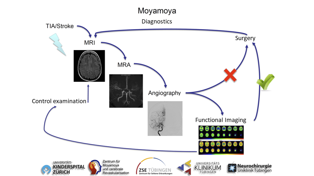
A detailed diagnostic work-up of patients is required for correct treatment decisions. These examinations are far more than conventional routine diagnostics and require a highly specialized center with sufficient expertise. Within many years of clinical experience and research activities, we have defined standardized diagnostic protocols for all patients to be able to define the therapeutic strategy. This ensures that patients / vascular territories get the treatment needed, either conservatively or surgically. Follow-up examinations after surgery, as well as in patients with conservative treatment are defined in specific protocols.

High resolution MR imaging
Besides routine stroke sequences, we acquire special high-resolution sequences on a modern 3 Tesla MRI, which are capable to depict the vessel wall with a resolution of less than one millimeter. Thus, a possible activity and a progression of the disease can be recognized and adapted to the further treatment and control strategy.

Cerebral angiography
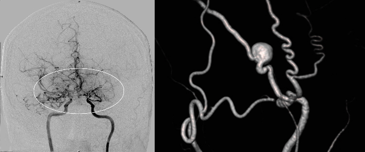
Conventional cerebral angiography is the most important diagnostic tool to diagnose Moyamoya disease. This examination can be compared to a cardiac catheterization, but for the cerebral arteries. In Moyamoya patients, as opposed to routine screening for other cerebral diseases, the anterior and posterior circulation of the brain is displayed selectively, as well as the supply of the extracranial vessels which might be needed as donor vessel or might build spontaneous EC-IC collaterals. This comprehensive examination is particularly important to understand the full extent of the disease and to understand any altered blood flow in the brain. Depending on the findings, the stenosis of cerebral arteries, as well as possible concomitant pathologic changes, are selectively displayed in 3-dimensional high-resolution angiography. It is important to mention that Moyamoya disease cannot and should not be treated by catheter intervention (stenting), as several studies have shown that this might lead to hemorrhage or rapid re-occlusion.

Functional MRI (breath-hold fMRI)
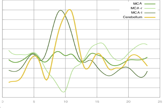
In conventional angiography and conventional MRI, the vessels, blood flow, and tissue can be displayed. However, these studies provide no information about the absolute blood flow and volume of the brain. MR and CT perfusion examinations of cerebral blood flow have been shown to be insufficiently sensitive to reliably depict the blood circulation in Moyamoya patients due to the existing collaterals. With intensive developments of functional MRI, we have established an imaging protocol that can depict the reactivity of the cerebral vessels (and thus indirectly the cerebral perfusion reserve). In the so-called “breath-hold” fMRI, patients have to hold their breath for several seconds in few cycles. This increases the CO2 concentration in the blood and cerebral arteries dilate. This change in blood flow can then be measured indirectly. Since this is a very new method, it must be mentioned that medical decisions are primarily exclusively in synopsis of all findings including the use of PET-CT. However, breath-hold fMRI has proven to be a good screening tool for long-term follow-up where it is mainly used.

H2 15O PET CT
The most important study for the examination of the blood flow in the brain at rest (baseline) and after drug-stimulated vessel dilation (“Diamox / acetazolamide challenge”) is the so-called water (H2 15O) PET CT. In this study, a defined amount of short-term radioactive water is applied to the blood system and thus the absolute amount of blood in the brain can be determined for each territory. This is then done both at rest and after medication. Thus, the cerebral perfusion reserve can be measured absolutely and analyzed based on each vascular territory. With this information a tailored revascularization strategy can be planned. As this is a very special nuclear-medicine examination, it can only be carried out in a few places with lots of experience with this technique.

