Etiology and Symptoms
The etiology of Moyamoya disease is still largely unknown. Genetics, environmental factors (e.g. radiation or meningitis) or local inflammation in the vessel wall are suspected to cause the disease. It is particularly important to distinguish Moyamoya from a classic cerebral vasculitis. A vasculitis can be diagnosed or excluded by a lumbar puncture and angiography also looks different than in Moyamoya. However, if the findings are inconclusive and changes primarily affect the basal cerebral arteries, Moyamoya disease must also be considered.
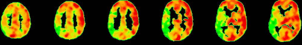
In Moyamoya patients symptoms are caused by a progressive narrowing of the blood vessels supplying the brain, which mainly affects the main cerebral arteries of the circle of Willis. Such stenosis is usually surrounded by newly developed fine collaterals, which are often insufficient. As result, neurological symptoms (headache, hemiplegia, seizure) due to a blood-shortage of the respective brain area might happen as transient ischemic attack (TIA) or manifest stroke (ischemic stroke). Frequently after the first appearance of symptoms patients recover well, but further diagnostics and possible therapy should be initiated quickly, in order to prevent permanent damage by more severe strokes. The pathophysiological causes of the symptoms are often TIAs, in which a transient insufficient supply of the brain leads to reversible neurological deficits. If a manifest stroke has occurred, the size and location of the stroke determines whether the symptoms are transient or permanent. More unspecific symptoms might be a general fatigue and decreased resilience, so that the head feels “like under a bell”, occasionally accompanied by dizziness.
Cerebral hemorrhage (hemorrhagic stroke) might occur due to the rupture of fragile fine surrounding collateral vessels, but is seen rarely (about 10%) in European patients. It can also lead to sudden neurological deficits and severe headaches or to a life-threatening unconsciousness.
After first the symptoms, detailed diagnostics and if necessary, referral to a specialized center is important, so that the severity of the disease can be evaluated precisely to initiate possible therapeutic steps.